NIG course for Zebrafish Imaging and Transgenesis was held at National Institute of Genetics (NIG) in Mishima, Japan during July 29-Aug 8, 2015. It was sponsored by NIG, and hosted by Koichi Kawakami’s lab. The first half of the course was devoted to CRISPR/Cas9-mediated DNA editing for mutagenesis and knock-in. In the second half, we performed calcium imaging of the zebrafish larval brain.
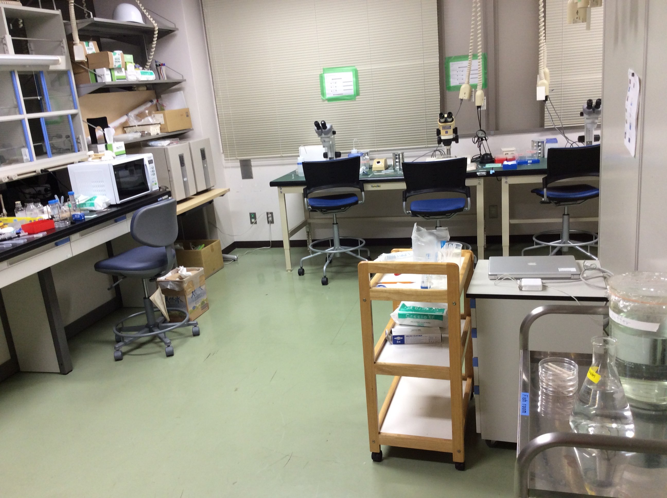
For the training course, we brought all the stuff needed into an empty room: lab equipment, five stereomicroscopes, and five epifluorescence microscopes equipped with Hamamatsu cameras.
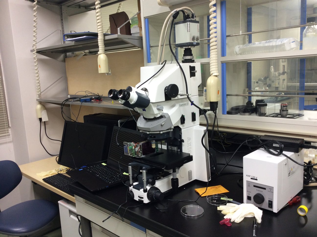
Microscope:ZEISS Examiner D.1, Objective lens: IR-ACHROPLAN 40x/0,80W 440095, Camera:HAMAMATSU ORCA-Flash4.0, Desktop PC:DELL Precision T5500, Laptop PC:FRONTIER, DAQ:NI USB-6001
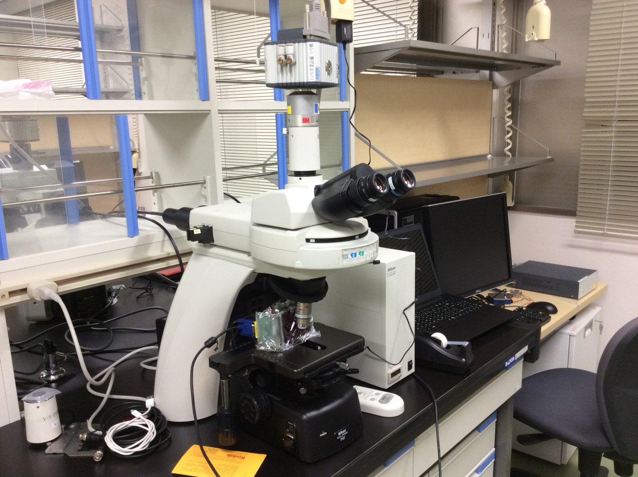
Microscope:NIKON, Camera:Hamamatsu ORCA-R2, Desktop PC:DELL Precision 5500, Laptop PC:FRONTIER, DAQ:NI USB-6001
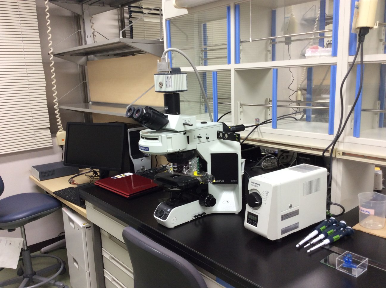
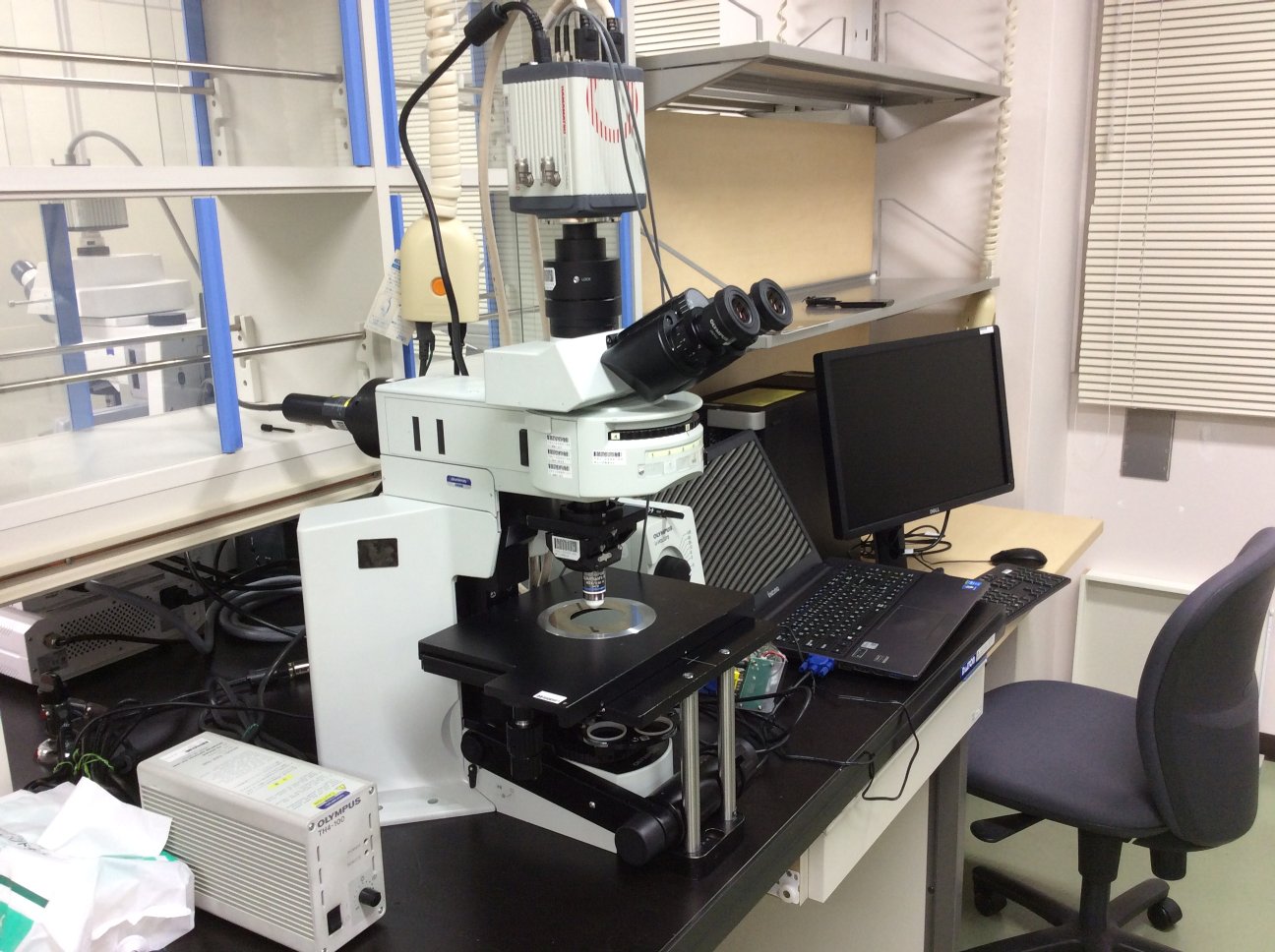
Microscope:OLYMPUS BX53, Camera:Hamamatsu ORCA-R2, Desktop PC:DELL Precision 5500, Laptop PC: NEC LaVie (Windows8), DAQ:NI USB-6001
Microscope: OLYMPUS BX51WI, Camera: HAMAMATSU ORCA-Flash4.0, Desktop PC:DELL Precision 5500, Laptop PC: IIYAMA, DAQ:NI USB-6001
Microscope: ZEISS Imager Z.2, Camera: HAMAMATSU ORCA-R2, Desktop PC:DELL Precision T5500, Laptop PC:FRONTIER, DAQ:NI USB-6001
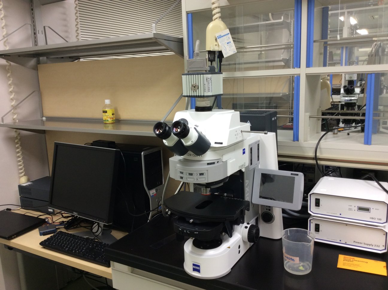
Image acquisition software: HCImage with High Speed Streaming module (ORCA-R2) or Hard Disk Record module (ORCA-Flash4.0) (Hamamatsu Photonics)
Software for visual stimulus presentation: Matlab R2015a with Psychtoolbox-3
We are very grateful to Hamamatsu Photonics, Zeiss, Olympus and Nikon for lending us the microscopes and cameras for this training course.
Web resources